genomics lab
About Our Services:
The award of the TransPOT grant has allowed us to acquire a state-of-the-art laser capture microdissection system and a robotic real-time PCR system for mid and high-throughput analyses. Trained personnel funded by TransPOT ensure the professional use of this equipment and aids users to optimize their experiments when using the facilities. Please feel free to contact us with regard to making an appointment to use the equipment.
The Laser Capture Microdissector (LCM) is able to isolate uncontaminated source material and allows isolation of homogenous analysis material. Because analyses of gene expression patterns rely on exactly-separated analytical material, unwanted cells may alter results and conceal the signals of the relevant cells. The LCM uses a focused laser beam to cut out and isolate a selected specimen without contact, rapidly and in an uncontaminated form. Objectives with a high numerical aperture bundle the energy so precisely that they are able to work to an accuracy of well under a micrometer and manipulation is possible down to the subcellular level, without involving neighbouring tissue. The laser pulse of the Laser Capture Microdissector only affects the specimen for approximately one nanosecond. In this short time heat transfer to adjacent tissue is negligible, ensuring gentle isolation that also allows for live cell recultivation. Isolatation of sensitive stem cell specimens is possible without influencing their vitality or genetic structure.
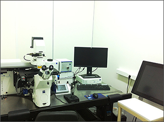
The LCM acquired is the PALM MicroBeam Microdissection system of Carl Zeiss MicroImaging GmbH, Germany, one of the most reputable companies of microscopy and imaging world-wide. The system acquired is unique in Greece and comprises:
- Reverse phase microscope AxioObserver Z1 with TFT touchscreen monitor. This feature can be used for the research of cell cultures in many biomedical applications, e.g. cell biology, pharmacology, molecular genetics, developmental biology and the neurosciences, and also for more basic research. The system has been specially designed for fluorescence applications and time-lapse experiments under live cell conditions. The supplied hardware comprises a sensor module on the nosepiece carrier, a beam combiner below the nosepiece and a Definite Focus controller. Definite Focus is coupled directly to the nosepiece carrier and directs infrared light of wavelength 835 nm into the beam path of the microscope. Definite Focus effectively counteracts drifting in the Z-direction and the microscopic image always remains in focus. This feature can be operated directly via the TFT touchscreen display of Axio Observer.Z1. Furthermore, preparation and waiting times in time-lapse experiments are markedly reduced. Convenient additional functions such as the storage of two nominal values and the possibility of using the system as a "focus finder" after replacement of the culture vessel are integrated into the system. Furthermore, Definite Focus is fully compatible with the AxioVision and LSM software from Carl Zeiss. The module supports all Axio Observer options such as TIRF, LSM or multiphoton microscopy. It works with all standard culture vessels, including multiwell dishes with glass or plastic bottoms.
- Optical equipment and multifluorescence channel (DAPI, FITC, CY3, GFP, rhodamine). Whatever the dye used, a simple change of filter results in the system automatically moving the sample grid to the correct position.
- Digital cameras. The technology of the equipment coverts the research microscope into a reliable, high precision laser tool, but it also can be used as a normal research microscope without any restrictions on the standard functions of light microscopy. Techniques such as phase contrast, PlasDIC or regular immunofluoresence analysis are all possible.
- Contact-free laser microdissector unit with micro-tweezers. Optical tweezers, also known as laser tweezers, are a highly effective and non-invasive method to manipulate cells as well as trap, move and sort microscopic particles. The selected specimen is separated with a software-controlled laser beam and transported by a laser pulse into a collection device.
- Robomover, enabling reliable and computer controlled collection devices of the PALM systems. The Robomover is the advanced form of the CapMover. It enables, for example, the exact division of several selected elements into different Caps or even in the recesses of a microtitre plate. The RoboMover automatically sorts the colour-coded elements into the pre-set collection devices so that hundreds of samples can be precisely sorted and collected.
- Incubator with software-controlled thermostat. This on-stage incubation enables temperature control in combination with CO2 and humidity. A heated lid stabilizes the temperature of the complete inner volume, so that no condensation can occur even under humid conditions.
- Apotome.2, which provides a tremendous improvement in image quality and is advantageous in 3D fluorescence microscopy. This system creates optical sections of fluorescent samples, free of scattered light. Apotome.2 can recognise the magnification and move the appropriate grid into the beam path. The system can then calculate the optical section from three images with different grid positions without time lag.
- Microinjection system (Eppendorf, Germany) for adherent cells. This feature can also be used with early stage embryos and small organisms. Semi automatic injection ensures high cell survival rates and the injection point and pressure of injection can be controlled.
- Computer and software. This system offers complete flexibility in observation, manipulation and analysis of cells. Developed in order to make even complex applications with living cells economical, the microscope software has high standards in user guidance, individualization and range of performance. AxioVision has the potential to grow as tasks dictate and its analysis modules offer the most technically advanced solution for almost any application.
The PALM Microbeam is suited to the workflow of even the most complex experiments and provides gentle handling of fluorescence specimens and living cells. It can be used on standard slides with no intermediate steps required to give reliable isolation and recultivation of living cells in a sterile environment.
The LCM may be used for the following applications: fixed tissue isolation (human, mouse and drosophila tissue), non-contact Laser Capture Microdissection (LCM) and recultivation of live cells; chromosome isolation and processing; subsequent isolation of DNA, RNA and protein for genomic or proteomic analyses. Examination of human blood and tissue samples, observation of intracellular
processes in living cell cultures, cell/cell interactions, motility, growth,
measurement of potentials, drug detection, microinjection, IVF (in-vitro
fertilization), toxicity studies, patch-clamp techniques, ion measurements,
digital recordings, time lapse studies with automated processes,
z-sectioning, deconvolution, visualization of molecular structures, Fura (Ca
measurement), GFP, laser capture microdissection, single molecule detection
etc.
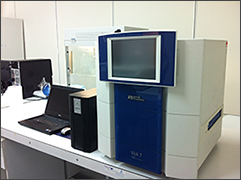 With a simplified workflow, intuitive software, touch-screen interface, and one-button protocols for error minimization, the ViiA™ 7 System RT-PCR engine has exceptional reproducibility with minimal well-to-well and instrument-to-instrument variation. Full compatibility with any standard or fast-cycling 384- or 96-well plates and TaqMan® Array Micro Fluidic Cards and Reagents makes high-productivity real-time PCR accessible to any sized lab.The Applied BiosystemsViiA 7 Real-Time PCR System uses fluorescent-based polymerase chain reaction (PCR) reagents to provide: With a simplified workflow, intuitive software, touch-screen interface, and one-button protocols for error minimization, the ViiA™ 7 System RT-PCR engine has exceptional reproducibility with minimal well-to-well and instrument-to-instrument variation. Full compatibility with any standard or fast-cycling 384- or 96-well plates and TaqMan® Array Micro Fluidic Cards and Reagents makes high-productivity real-time PCR accessible to any sized lab.The Applied BiosystemsViiA 7 Real-Time PCR System uses fluorescent-based polymerase chain reaction (PCR) reagents to provide:
- Quantitative research detection of target nucleic acid sequences using real-time analysis.
- Qualitative research detection of targets using post-PCR analysis.
- Qualitative analysis of PCR product achieved by melt culve analysis that takes place post-PCR.
It has also been designed to improve researcher productivity for research. The system optimizes and simplifies a host of real-time PCR applications; all integrated and easily accessible from a touch screen. OptiFlex™ System within the ViiA 7 provides enhanced fluorescence detection.Maximum multiplexing: Six decoupled excitation and emission filter channels for the greatest number of dye combinations and maximum multiplexing capabilities.
- Flexible data collection: Multiple ramp method detection formats provide more flexibility for collecting data during a ramp stage.
- Precise quantification: Detect as small as 1.5–fold changes in target quantities in singleplex reactions.
|